Electromyostimulation, an effective, classic therapeutic tool that is still little used because it is little known
About the author: Docteur P.Jenoure, Ars Ortopedica, Gravesano
Introduction
While many people are probably aware of the primordial role of muscle in generating movement in the various segments of the body, far fewer are aware of the formidable stabilizing, restraining and cushioning capacities of this same muscle tissue. But it turns out that these faculties, remarkable though they are, are put to the test in the context of high-intensity or repetitive demands, as encountered in professional life as well as in leisure activities, particularly sport. That’s why it’s a good idea to look after your muscles, and if necessary, use proven auxiliary maintenance techniques.
Musculature
Contrary to the humorists who have a field day with this theme, it seems incongruous to us to want to establish a hierarchy among the organs of the human body, so remarkable is the functional interdependence between its different parts. Nevertheless, striated muscle tissue, with its 600+ muscles representing between 45% and 55% of total body mass, containing around 50% of the body’s total proteins, and above all, playing essential and vital roles, is an extremely important tissue. It’s important to remember that, in addition to its mechanical function, the musculature produces heat and energy to keep body temperature constant, serves as a storage site for amino acids, lipids and carbohydrates, helps maintain a basic metabolism and plays a quasi-endocrine role in blood sugar regulation.
A striated skeletal muscle is made up of muscle fibers, giant cells that are very elongated, striated in appearance and electrically excitable. Each muscle fiber has the ability to shorten following contraction triggered by a nerve stimulus arriving at a motor plate. This shortening takes place within an even smaller unit, the myofibril, made up of actin and myosin, protein filaments arranged in sarcomeres, interlocking and sliding one inside the other. It thus forms a linear driving element capable of delivering motion and force. However, this description is too general, and muscle tissue is not a homogeneous structure. In fact, it’s made up of different types of muscle fibers with different histological, metabolic and contractile properties, adapted to their specific area of functional activity. There are 3 main types: type I, type IIa and type IIb fibers. I-fibres are also known as slow or tonic fibres, and produce contractions of low amplitude, but can do so for prolonged periods. IIa fibers, fast or tonico-phasic fibers, produce strong contractions, but of shorter duration than I fibers. Finally, IIb fibers, also fast, or explosive, produce very powerful but brief contractions. The distribution of each type in the musculature is genetically fixed, and in the normal population, the proportion of I and II fibers is more or less equal. However, sports medicine research has shown that, depending on the sporting specialization, this distribution can change significantly: some sprinters, for example, have a proportion of fast type II fibers of 70%. Another amazing property is the ability of fibers to transform according to the type of training (I→II, or II→I).
Another key point in relation to electrostimulation is the notion of motor unit. This is the entity formed by a medullary alpha motor neuron of a given type and all the muscle fibers of the same type that it innervates. One complication is that each fiber type has a different range of stimulation frequencies: from 8 to 30 Hz for type I, from 20 to 50 Hz for type IIa, and from 30 to around 65 Hz for type IIb.
So it’s easy to understand that effective training of a muscle as a whole absolutely requires the use of all the stimulation frequencies specific to each type of fiber. During a maximum voluntary contraction, all the motor units of the different types of fibers present in the muscle are called upon at their optimal frequency, enabling the development of maximum force. On the other hand, during a submaximal contraction, voluntary stimulation is “asynchronous”, which means that some motor units are recruited, while others are left at rest, and a kind of rotation between the units is established to protect against fatigue, which occurs more rapidly when the contraction must be maximal and all the units function “synchronously”.
Training
From a biological point of view, training, as we know it from sporting activities, is a set of stimuli to an organ or system of organs, stimuli that meet fairly well-defined criteria, and provoke disturbances at the cellular level of these targets that are not harmful but are sufficiently significant to induce adaptation mechanisms. This adaptation can be seen as a protective reaction. A simplistic but evocative example is given by the skin’s reactions when stimulated by ultraviolet radiation: if this is insufficient, there’s no change in complexion; if it’s too strong (in duration or intensity, or both), there’s sunburn, whereas with adequate exposure, the desired tan appears. A large number of organs react in this way – most of them, one might say, even those deemed “bradytrophic”: tendons, bones, cartilage, among others. The heart’s adaptations are fairly well known, and other “trainable” systems are regularly being discovered. But once again, muscle has some impressive features to offer. Just think of the changes visible to the naked eye in the external morphology of body-builders.
As stated in the introduction, inhomogeneous muscle tissue reacts inhomogeneously to stimuli. Endurance training (aerobic capacity) will generate responses favoring oxygen utilization (mitochondria), while so-called resistance efforts (anaerobic capacity) will promote other adaptations. In rehabilitation, the focus is on muscle hypertrophy, which is the main guarantee of an increase in strength capable of playing the important mechanical role in the “solidity” of the musculoskeletal system. To achieve this, we use intense efforts close to the muscle’s maximum strength (maximum strength = heaviest weight that can be mobilized during a single repetition by muscular contraction). But all these forms of stimulation have their uses, as they all contribute, to a greater or lesser extent, to improved vascularization, optimized metabolism and improved inter- and intramuscular coordination.
Training for maximum strength, which contributes to fiber thickening (hypertrophy) by mobilizing satellite cells, requires exhausting work that is difficult to achieve in the event of injury or illness.
Malfunctions
This organ, whose importance we emphasize, is obviously not immune to disorders of various kinds, which can manifest themselves as momentary or lasting deficiencies in voluntary activity. Today, it’s a relatively well-known fact that such deficiencies are accompanied fairly rapidly by a whole host of adverse effects: muscular atrophy, joint degeneration, disruptions to neural control, disturbances to blood circulation and trophicity, and loss of bone substance, to name but the main ones. It’s the image of a true maladjustment syndrome.
Muscular atrophy (amyotrophy) (1)
Muscular atrophy, or amyotrophy, is defined as a reduction in the mass of a muscle or muscle group, of various origins. First and foremost, this atrophy results from under-utilization, which may be the consequence of a neurological or bone problem, or of the muscle itself, or even of a metabolic disease. Probably the most frequent cause of atrophy is trauma to the musculoskeletal system (joint or muscle itself), and it’s not impossible that in such situations, the loss of muscle substance is a kind of protective reaction, since the resulting loss of strength inevitably prevents extreme movements likely to aggravate the injury.
This inevitable loss of strength when atrophy appears is not, however, a reliable clinical criterion, and for a serious determination, only the measurement of muscle mass by DPX or the measurement of the section slice by CT-scan, sonography or MRI are scientifically rigorous methods.
Today, the phenomenon of atrophy is partially understood through the subtle insights of molecular biology, but more in-depth knowledge will be needed to develop more effective rehabilitation treatments or pharmacological interventions.
Sarcopenia (2,3,4,5,6,7,8)
All individual variations considered, peak muscle mass is reached around the age of 30, then declines by approximately 5% per year from the age of 40 onwards, with an accelerated rate of decline after the age of 60. This involuntary, quasi-physiological loss of muscle mass with age is the definition of sarcopenia, at least in broad terms. The prevalence of sarcopenia is of the order of 30% in the population over the age of 65, rising to 50% over the age of 80. This decline in muscle mass with age is more frequent and tends to be more severe in women.
Sarcopenia is obviously associated with functional decline (strength among other things), linked to an increased risk of falls, greater vulnerability to trauma, all of which lead to dependency.
The pathophysiology of this condition is fairly well understood today.
Various sarcopenia prevention and treatment programs have been studied, and while none of the approaches appears superior to the others, the positive role of strength training is regularly emphasized.
Electromyostimulation (9)
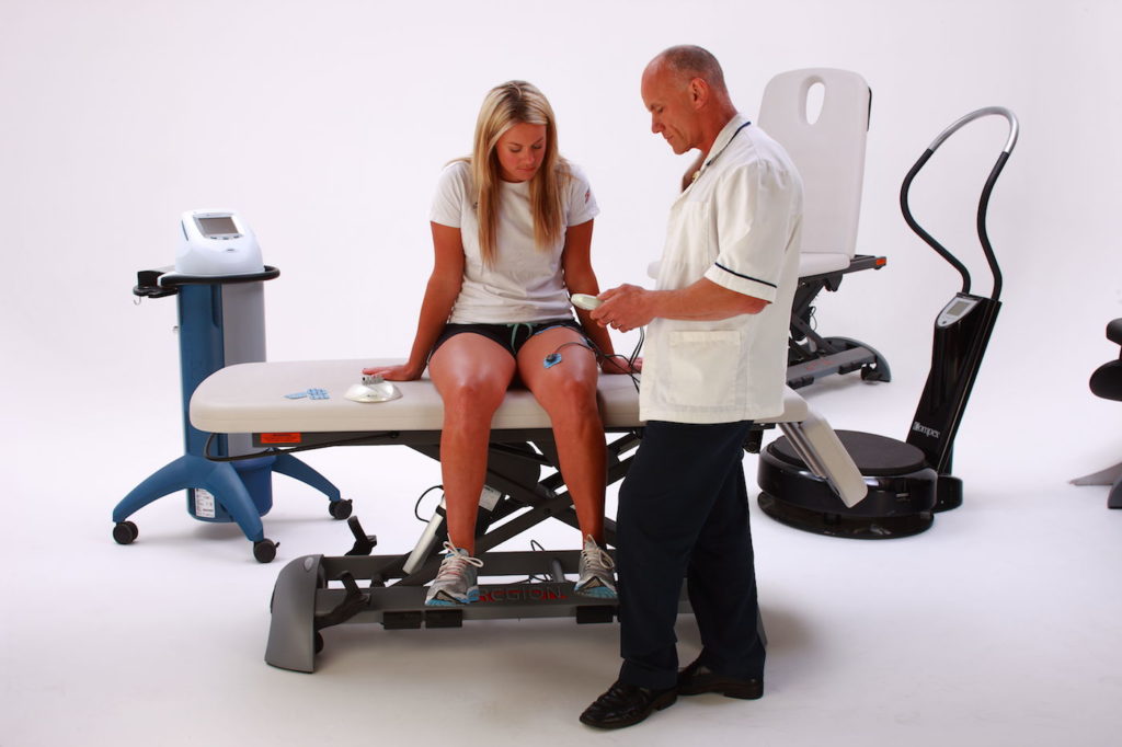
Electrostimulation is a technique that transmits, via the motor nerve, a variable electrical impulse (duration and intensity) from an electrical stimulator to selected muscles via two or more electrodes. The electrical impulse causes muscular contraction of the selected muscle(s) without involving the central nervous system (brain).
Electrostimulation has been used for many years by physiotherapists in post-operative rehabilitation or in treatments where the aim is to combat the risk of atrophy or to restore the musculature to a functional state. It is also used for sports preparation and recovery.
Electrostimulation, also known as electromyostimulation, is an ancient technique which has nevertheless undergone significant development thanks to the advent of new technologies.
Its essential purpose is to supplement or even replace a momentary or lasting deficiency in voluntary activity. But to achieve this, it has to mimic the natural functioning of muscle contraction as closely as possible. In view of this subtle functioning, the overall training required will absolutely have to call upon all physiological stimulation frequencies, as a single frequency, even if chosen within the range of natural frequencies, cannot achieve this objective of globality. But transcutaneous electrical stimulation is unable to discriminate in any way, and always provokes a “synchronous” or simultaneous response from all recruited muscle fibers. It systematically imposes the same frequency on all fibers without taking into account their own specificity. Consequently, to achieve effective overall training of the various types of fibers making up a given muscle, it will be necessary to perform a harmonious sequence of different stimulations with the appropriate frequencies, corresponding to the different types of fibers. And consider the essential recovery phases. Electromyostimulation training, technically very easy today, according to the concept defined, therefore requires quite a long time and regular repetition. Gone are the days of traditional 30-minute sessions, generally twice-weekly, with empirical stimulation parameters. Today, it’s all about long-term electromyostimulation, with automatic progressive sequencing of different, specific activity phases, which has scientifically demonstrated its effectiveness. This form of rehabilitation has many advantages: it is possible even when the general state or a particular situation does not allow effective activation of the muscles, for reasons of pain or disruption of motor control, for example. What’s more, it’s almost independent of strong motivation, unlike conventional training, which requires a great deal of discipline. In this context, the combination of electromyostimulation and physical training is particularly favourable. (10, 11). The combination of electrical stimulation and voluntary work may be of interest as the patient progresses, particularly to facilitate the restoration of good motor control.
Electromyostimulation applications
It therefore seems logical to use electromyostimulation as described in all situations where loss of muscle mass represents a handicap for the function concerned. That said, the field of application is very broad, and practically any atrophied muscle can expect to benefit from this proven but little-known rehabilitation technique (12,13).
The technique of muscular electrostimulation is well known in specialist circles, and many applications have been described.
The best-known is certainly the fight against quadricipital atrophy after knee surgery, in particular ligamentoplasty of the anterior cruciate ligament. But with the advent of conservative treatment of this damaged ACL, focused on optimizing the thigh muscle envelope, an excellent indication for electrostimulation is almost self-evident. (14,15, 16, 17, 18, 19, 20,21)
Another frequent and difficult-to-treat knee condition is anterior knee pain. Improvement of the vastus medialis, and especially its oblique component, is a classic, and EMS has proved effective in this situation too, as it enables analytical stimulation of this muscle head. It’s worth noting that some authors are now advocating hip rotator training in this very annoying condition, in order to improve the frequent malalignment observed in this clinical picture. Here too, electrostimulation can help.
In general traumatology, but in sports in particular, supination sprains of the tibio-tarsal joint are by far the most frequent accident (1 incident of this type per day for every 10,000 inhabitants in our type of society, i.e. 800 sprains every day for little Switzerland, 290,000 per year!) Sideration of the peroneal muscles during abrupt stretching and their concomitant dysfunction (temporal deactivation) can represent a major disturbing factor during rehabilitation. Appropriate electrostimulation has been shown to have a positive effect.
The frequency of back pain is quantitatively of a similar order of magnitude, and stimulation of the pelvic girdle muscles is one of the classic treatments, via active physiotherapy. Electromyostimulation has also proved effective in this situation (22).
The importance of the rotator cuff muscles in the proper functioning of the shoulder is well known. Electromyostimulation can be successfully applied in the case of overload lesions, dysbalance or after injury to these structures (23, 24).
By analogy, electrostimulation has been very successfully applied in rehabilitation after hip (25) and knee (26) prosthetic surgery, after osteotomies, in the treatment of fracture sequelae, and after tendon surgery, such as Achilles tendon suture, which is typically accompanied by significant calf atrophy.
In the case of various muscular contractures (spine, limbs), EMS has proved its worth.
We are also aware of satisfactory applications to counteract the after-effects of stroke, such as paresis and spasticity.
Finally, for the sake of completeness, we should mention the use of electrical muscle stimulation for urinary or anal incontinence, chronic cachectizing diseases (COPD, heart failure, cancer) (27, 28, 29,30) and, more recently, in combination with cybertheses to help rehabilitate paraplegic patients (31).
Practical aspects
The use of electromyostimulation should be linked to a medical prescription, possibly “inspired” by a physiotherapist. Under these conditions, the rental of the stimulation unit will be covered by insurance (accident insurance, health insurance). In order to achieve satisfactory efficacy, it is essential for the patient to have the device available at all times, since it will be used on a regular, daily basis, for at least a few weeks. With some suppliers, the device will be supplied programmed to the patient’s specific needs. The physiotherapist will determine the optimal location for the electrodes and mark the spot with a dermograph. It is he who will instruct the patient in correct use. It’s worth noting that some devices record all the “work” done, which is a good way of improving compliance. It’s also an excellent means of control for the prescriber, and if need be, for the insurer. Not to mention the scientific aspect.
Some devices are able to deliver analgesic currents, such as TENS, a useful complement in many situations.
Conclusions
Muscle tissue, which in the final analysis can be compared to an organ, is a rather special entity with numerous characteristics: unique quantitative importance in the human body, functional versatility that is equally unparalleled, remarkable plasticity, unfortunately in both the ana- and catabolic sense. Finally, in our context, we’d like to mention a high degree of sensitivity to the absence of normal neurological stimulation, manifested by rapid and often significant atrophy with deleterious consequences, in our view not always taken into account by medicine and doctors. Electromyostimulation is an effective and ultimately easy-to-use way of combating this condition.
Bibliography
1) Léger B, C.Gobelet (2011)
Muscular atrophy
Swiss Journal of Sports Medicine and Traumatology 59 (1), 14 – 17, 2011
2) Michel J.P, P.O. Lang, A.J Cruz-Jentoft (2009)
Sarcopenia: a new hot topic in geriatrics
Swiss Medical Journal, 2009, 5 :2200 -4
3) Lang P.O (2012)
Sarcopenia
Rheuma Switzerland, 1, 2012, 13 -15
4) Münzer T (2010)
Sarcopenia in the elderly. Concept, clinic and interventions
Forum Med Suisse, 2010; 10 (10): 188 -190
5) Janssen I, (2006)
Influence of sarcopenia on the development of physical disability: The cardiovascular Health Study.
J Am Geriatr Soc, 2006. 54: p. 56-62.
6) Orr R, Raymond J, Fiatarone Singh M. (2008)
Efficacy of progressive resistance training on balance performance in older adults: a systematic review of randomized controlled
trials.
Sports Med. 2008;38(4):317-43
7) Talbot L.A, Gaines J.M, Ling S.M, Metter E.J (2003)
A Home-Based protocol of Electrical Muscle Stimulation for Quadriceps Muscle Strength in Older
Adults with Osteoarthritis of the Knee
J Rheumatol, 30: 1571-157
8) Caggiano E., Emrey T., Shirley S., Craik R.L. (1994)
Effects of electrical stimulation or voluntary contraction for strengthening the quadriceps femoris muscle in an aged male population .
Journal of Orthopaedic and Sports Physical Therapy, 20, 22-28
9) Brodard R.
A fundamentally innovative scientific concept finally makes electrostimulation muscle training optimally effective
Personal communication
10) Paillard T
Combined Application of Neuromuscular Electrical Stimulation and Voluntary Muscular Contractions
Sports Med 2008; 38 (2): 161-177
11) Wigerstad-Lossing I, Grimby G, Jonsson T., Morelli B., Peterson L., Renström P.(1988)
Effects of electrical muscle stimulation combined with voluntary contractions after knee ligament surgery.
Medicine and Science in Sport and Exercise, 20, 93-98
12) Qin L., Appell H.J., Chan K.M., Maffulli N. (1997)
Electrical stimulation prevents immobilization atrophy in skeletal muscle of rabbits .
Arch. Phys. Med. Rehab, Vol. 78, 512-17
13) Valli, P. Boldrini, L., Bianchedi, D. and co (2002)
Effects of low intensity electrical stimulation on quadriceps muscle voluntary contraction
Journal of Sports Medicine and physical fitness, 42: 425-430
14) Arvidsson I., Arvidsson H., Eriksson E., Jansson E. (1986)
Prevention of quadriceps wasting after immobilization: an evaluation of the effect of electrical stimulation .
15) 0’Connor J.J. (1993)
Can muscle co-contraction protect knee ligament after injury or repair?
British Editorial Society of Bone and Joint Surgery, 75-B,41-48
16) Kain C.C., McCarthy J.A., Arms S., Pope M.H., Steadman J.R., Manske P.R., Shively R.A. (1988)
An in vivo analysis of the effects of transcutaneus electrical stimulation of the quadriceps and hamstrings on anterior cruciate ligament deformation .
The American Journal of Sports Medicine, Vol. 16, No 2, 147-152
17) Snyder-Mackler L., Ladin Z., Schepsis A.A., Youg J.C. (1991)
Electrical stimulation of the thigh muscles after reconstruction of the anterior cruciate ligament .
The Journal of Bone and Joint Surgery, 73-A, 1025-36
18) Nitz A.J., Dobner J.J. (1987)
High intensity electrical stimulation effect on thigh musculature during immobilization for knee sprain .
Physical Therapy, Vol. 67, No2, 219-22
19) Delitto A., McKowen J.M., McCarthy J.A., Shively R.A., Rose S.J. (1988)
Electrically elicited co-contraction of thigh musculature after anterior cruciate ligament surgery .
Physical Therapy, Vol. 68, No1, 45-50
20) Morrissey M.C., Brewster C.E., Shields C.L., Brown M. (1985)
The effects of electrical stimulation on the quadriceps during postoperative knee immobilization .
The American Journal of Sports Medicine, Vol. 13, No1, 40-45
21) Gould N., Donnermeyer D., Gammon G.G., Pope M., Ashikaga T. (1982)
Transcutaneous muscle stimulation to retard disuse atrophy after open meniscectomy.
Clinical Orthopaedics and Related Research, No178, 190-197
22) Coghlan S, L.Crowe, U. McCarthy Persson, C. Minogue, B. Caulfield
Neuromuscular Electrical Stimulation Training Results in Enhanced Activation of Spinal Satabilizing Muscles During Spinal Loading and Improvement in Pain Ratings
23) Reinold M.M, L. C Macrina, K. E. Wilk, J. R.Dugas, E.L.Cain ,J.R.Andrews (2008)
The effect of neuromuscular electrical stimulation of the infraspinatus on shoulder external rotation force production after rotator cuff repair surgery
Am. J. Sports Med. 2008; 36; 2317
24) Colson S.S, M. Benchortane, V. Tanant, J.P. Faghan, M. Fournier-Mehouas,
C. Benaïm, C.Desnuelle (2010)
Neuromuscular Electric Muscular Stimulation Training:A safe and effective Training for Fascioscapulohumeral Muscular Dystrophy Patients
Archives of Physical Medicine and Rehabilitation, Vol 91, No5, May 2010
25) Gremeaux V, Renault J, Pardon L, Deley G, Lepers R, Casillas J-M (2008)
Low-frequency electric muscle stimulation combined with physical therapy after total hip arthroplasty for hip osteoarthritis in elderly patients: a randomized controlled trial
Arch Phys Med Rehabil 2008;89:2265-73
26) Avramidis K, Strike PW, Taylor P N, Swain I D (2003)
Effectiveness of electrostimulation of the vastus medialis muscle in the rehabilitation of patients after total knee arthroplasty
Arch Phys Med Rehab, 84: 1850-1853
27) Zanotti E, Felicetti G, Maini M, Fracchia C (2003)
Peripheral muscle strength training in bed-bound patients with COPD receiving mechanical ventilation
Chest, 124: 292-296
28) A. Abdellaoui, C. Pre’faut, F. Gouzi,, A. Couillard, M. Coisy-Quivy, G. Hugon,
N. Molinari, T. Lafontaine, O. Jonquete, D. Laoudj-Chenivesse and M. Hayot:
Skeletal muscle effects of electrostimulation after COPD exacerbation: a pilot study
Eur Respir J 2011; 38: 781-788
29) Lara Maris Napolis, Simone Dal Corso, Jose’ Alberto Neder, Carla Malaguti,
Ana Cristina Oliveira Gimenes, Luiz Eduardo NeryI
Neuromuscular electrical stimulation improves exercise tolerance in chronic obstructive pulmonary disease patients with better-preserved fat-free mass
CLINICS 2011; 66(3):401-406I
30)Isabelle VIVODTZEV, Richard DEBIGARÉ, Philippe GAGNON, Vincent MAINGUY, Didier SAEY, Annie DUBÉ, Marie-Ève PARÉ, Marthe BÉLANGER , François MALTAIS Functional and muscular effects of neuromuscular electrical stimulation in patients with severe COPD: a randomized clinical trial
Chest; Prepublished online November 23, 2011;
31) www.fscsfc.org/index.php?option=com_content&view=article&id=46&Itemid=2&lang=fr
Despite more than 30 years of personal contact with various representatives of one of the first neuromuscular stimulation systems, the author claims to have no conflict of interest.
Dr.med. P.Jenoure
ARS Ortopedica
ARS Medica Clinic
6929 Gravesano / Lugano